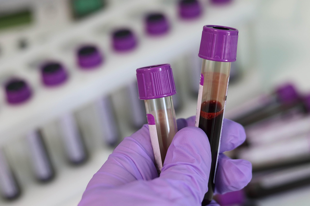
Patients with malignant diseases undergo various procedures to confirm the diagnosis, evaluate the extent of the disease, and assess therapeutic efficacy. One such procedure is tissue biopsy – sampling of cells or tissue for microscopic analysis. However, the very notion of biopsy often evokes fear and discomfort, leading to the increased use of its non-invasive alternative – liquid biopsy. This method has revolutionized clinical oncology by enabling simple tumor sampling, repeated molecular assessments, the design of tailored therapeutic strategies, and screening for therapeutic resistance.
What is a Liquid Biopsy?
The concept of personalized oncology is increasingly emphasized as an innovative approach built upon the ability to sequence each patient’s tumor. Its goal is to individualize therapy and monitor tumor evolution during specific treatment. Tissue biopsy remains the “gold standard” for diagnosis and molecular characterization. However, its limitations are substantial, including procedural invasiveness and potential complications such as bleeding, injury, infection, pain, limited tissue yield, and tumor heterogeneity. Consequently, liquid biopsy has emerged as a valuable, non-invasive, repeatable diagnostic method capable of profiling the molecular landscape of individual tumors and guiding clinical decision-making in cancer care.
Liquid biopsy is a diagnostic procedure that includes analysis of circulating tumor cells (CTCs) or tumor-derived DNA fragments from a blood sample. This approach can be utilized for early cancer detection, treatment planning, evaluation of therapeutic effectiveness, and surveillance for disease recurrence. Its major advantage is the ability to frequently sample blood over time, allowing clinicians to monitor dynamic molecular changes within the tumor. The rapid expansion of liquid biopsy in oncology is driven by advancements in comprehensive genomic profiling assays and evidence from numerous preclinical and clinical studies showing its clinical utility.
What can be analysed?
The tumor microenvironment comprises diverse cellular and non-cellular components, many of which originate from tumor cells and contribute to cancer cell survival, invasiveness, metastasis, and treatment resistance. Among these components, circulating tumor cells (CTCs), circulating tumor DNA (ctDNA), cell-free DNA (cfDNA), and extracellular vesicles, precisely exosomes, have emerged as valuable biomarkers detectable in the peripheral blood of oncology patients. Blood samples, including plasma, serum, or cellular fractions, can be analysed to detect and characterize multiple circulating biomarkers in detail.
These biomarkers can also be found in various other body fluids, expanding the reach of liquid biopsy beyond peripheral blood. They have been detected in prostatic secretions, urine (especially in renal, bladder, and prostate cancers), cerebrospinal fluid (particularly in central nervous system tumors and metastases), saliva and sputum, pleural effusions (notably in head and neck cancers and lung cancer), tears, bile, peritoneal washings, bone marrow (especially in breast, prostate, and lung cancers), stool, breast milk and ascitic fluid.
CTCs, ctDNA, and Exosomes
CTCs are malignant cells that shed into the bloodstream from primary tumors or metastatic sites at any stage of tumorigenesis. They circulate as single cells or as clusters, the latter has greater metastatic potential. CTC levels fluctuate dynamically and more accurately reflect tumor status than conventional blood-based markers. A reduced CTC count has been associated with improved overall survival, particularly in breast cancer. Additionally, CTCs show potential for early detection of some cancers such as lung cancer, although evidence is obtained from small cohorts of patients with chronic obstructive pulmonary disease. Diagnostic relevance has been further supported by subsequent findings of pulmonary nodules and confirmatory histopathology.
DNA is released into circulation in fragments (cfDNA) through apoptosis, necrosis, and active secretion by normal cells (notably lymphoid and myeloid cells due to rapid hematopoietic turnover) and malignant cells (ctDNA). Owing to its tumor-derived origin, ctDNA provides information on point mutations, copy-number variations, structural rearrangements, loss of heterozygosity, gene fusions, methylation changes, integrated oncogenic viral sequences, and other genomic signatures. Studies have shown that ctDNA offers a more comprehensive view of tumor characteristics and evolution, encompassing alterations originating from primary and metastatic lesions. Furthermore, mutations undetectable in conventional tissue biopsies may be identified through ctDNA analysis obtained via liquid biopsy.
Tumor-derived exosomes are small (30–120 nm), spherical vesicles surrounded by a lipid bilayer and containing proteins, DNA, RNA, small non-coding RNAs such as microRNAs (miRNAs), and lipids. Tumor cells actively secrete exosomes and use them for intercellular communication between the tumor and various cell types. Because each tumor cell releases a unique exosomal profile, exosomes may help in identifying the presence and type of malignancy. Their high abundance across biological fluids further enhances detection sensitivity. However, the lack of standardized, reliable isolation methods and analytical procedures remains a significant barrier to their widespread clinical implementation.
Clinical Potential and Challenges of Liquid Biopsy
New evidence confirms that liquid biopsy is a minimally invasive, high-value diagnostic tool enabling early tumor detection, real-time monitoring of treatment response, screening of high-risk individuals, assessment of tumor heterogeneity, and identification of emerging mutations. Similar to tissue biopsy, it provides detailed molecular information and can reveal tumor-related changes long before conventional diagnostic methods. By capturing intratumoral heterogeneity and clonal evolution, liquid biopsy enhances understanding of disease dynamics and may reveal occult metastases, potentially reducing recurrence risk.
Despite its advantages, broader clinical adoption remains constrained by limited sensitivity and specificity, insufficient methodological standardization, and high costs. Continued technological advances are essential for improved tumor detection across organ systems and for deeper molecular characterization, ultimately enabling more comprehensive integration of liquid biopsy into routine clinical practice.
Translated by: Nina Mikulić
Literature
2. Lone SN et al. Liquid biopsy: a step closer to transform diagnosis, prognosis and future of cancer treatments. Mol Cancer, 2022, 21, 79.
3. Caputo V et al. Diagnostic value of liquid biopsy in the era of precision medicine: 10 years of clinical evidence in cancer. Explor Target Antitumor Ther, 2023, 4(1), 102-138.